
Calabasas, California – October 01, 2026 – PRESSADVANTAGE –
Micro Medical Devices (MMD), a developer of portable ophthalmic diagnostic technologies, announced findings from an observational study evaluating the VF2000-NEO for visual field assessment in glaucoma care.
The study compared VR-based perimetry with conventional stationary perimetry and examined how visual field measurements from each method correlated with retinal structure measurements obtained through optical coherence tomography (OCT).
The study, “Comparison of VR-Based and Conventional Perimetry in Relation to OCT-Derived Structural Measurements,” was presented at the 17th European Glaucoma Society Congress, held May 30–June 2, 2026, in Brussels.
The findings are relevant to the assessment and monitoring of open-angle glaucoma, where clinicians routinely examine the relationship between functional visual field loss and structural changes to the retinal nerve fiber layer and ganglion cell complex. However, the results should be interpreted within the context of the study’s observational design and the smaller subset of patients who met the researchers’ high-reliability criteria.
“This study provides encouraging evidence that VF2000 visual field measurements can correspond with structural findings on OCT,” said Ramin Hooriani, President and Head of Software Development at Micro Medical Devices. “Understanding the relationship between structural and functional measurements is important in glaucoma assessment. These findings warrant continued evaluation of portable visual field testing technologies and their potential role in clinical practice.”
The study included 97 patients who underwent visual field testing with both the MMD VF2000-NEO and a conventional stationary Optopol perimeter. Each patient also received OCT imaging during the same visit.
Researchers compared functional visual field measurements—including mean deviation and pattern standard deviation—with two OCT-derived structural measurements: retinal nerve fiber layer thickness (RNFL) and ganglion cell complex thickness (GCC).
Thinning of the RNFL and GCC can indicate structural damage associated with glaucoma, while visual field testing measures corresponding changes in visual function. Evaluating both forms of testing can help clinicians assess whether structural changes observed through OCT align with a patient’s functional vision loss.
Of the 97 patients enrolled in the study, 38 met the study’s high-reliability criteria and were included in the high-reliability correlation analysis. Because this analysis represents a subset of the total study population, the findings should be interpreted cautiously and may not be generalizable to all patients or clinical settings.
Within this 38-patient high-reliability subset, the VF2000-NEO showed numerically stronger structure-function correlations than the conventional stationary perimeter across the four reported comparisons. Mean Deviation versus RNFL correlation was 0.40 for the VF2000-NEO compared with 0.30 for the conventional perimeter; Mean Deviation versus GCC was 0.55 compared with 0.50; Pattern Standard Deviation versus RNFL was -0.35 compared with -0.27; and Pattern Standard Deviation versus GCC was -0.60 compared with -0.50.
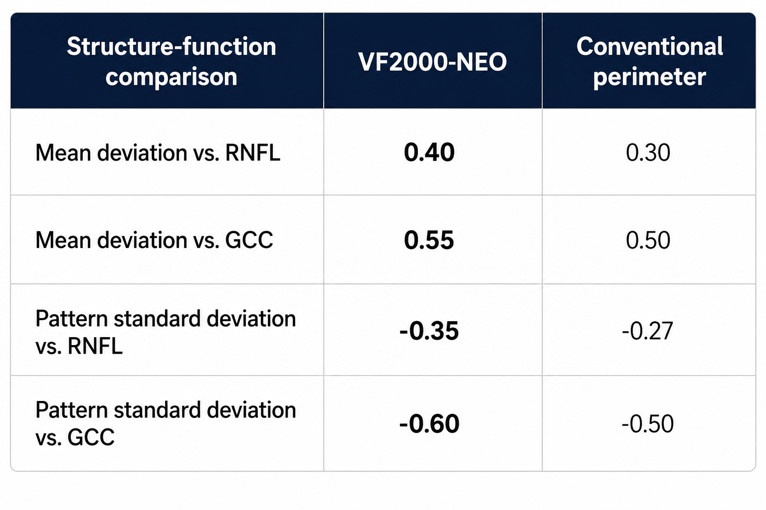
The correlation between mean deviation and RNFL thickness reached statistical significance for the VF2000-NEO but did not reach statistical significance for the conventional perimeter. Statistical significance in one comparison should not be interpreted on its own as demonstrating superiority of one testing platform over another.
Taken together, the observed results indicate that, among patients meeting the study’s high-reliability criteria, VF2000 functional measurements were associated with structural retinal measurements identified through OCT. Additional research involving larger patient populations and independent validation would help determine how broadly these findings apply to routine glaucoma assessment.
Open-angle glaucoma can progress gradually and without noticeable symptoms during its earlier stages. Clinical assessment therefore commonly includes both structural testing, such as OCT, and functional testing, such as visual field perimetry.
In patients with lower visual field test reliability, the researchers reported that VR-based perimetry was non-inferior to the conventional stationary perimeter. [Insert the study’s predefined non-inferiority margin, confidence interval and applicable statistical result, if available.] Among patients with high test reliability, the VF2000 produced numerically stronger correlations with OCT structural measurements in the comparisons reported by the researchers.
The researchers concluded that VR-based perimetry may provide robust functional-structural associations under reliable testing conditions. These findings reflect the results of this observational study and should not be interpreted as establishing broader clinical superiority or equivalence of the VF2000-NEO across all patients, testing environments, or glaucoma-care applications.
In addition to evaluating correlations with OCT measurements, the study compared examination times for the two testing methods.
The reported examination times were approximately 2 minutes and 50 seconds per eye for the VF2000-NEO and approximately 5 minutes and 20 seconds per eye for the conventional perimeter, representing an examination-time reduction of approximately 47% in this study.
The observed difference in testing time suggests that portable VR-based perimetry may offer workflow advantages in some clinical environments. However, examination time can be influenced by patient characteristics, testing protocols, operator procedures, and other factors, and the results of this study should not be assumed to apply to every clinical setting.
“The results suggest that portable visual field testing can combine clinically relevant measurements with shorter examination times under the conditions evaluated in this study,” Hooriani said. “We believe these findings provide a strong basis for continued clinical research into how portable perimetry can be incorporated into glaucoma care.”
Hooriani leads MMD’s software development and product innovation initiatives. He earned a Bachelor of Science in Electrical Engineering and Applied Physics and a Master of Science in Digital Signal Processing from California State University, Northridge.
The findings were also a topic of discussion during MMD’s participation in the 2026 World Ophthalmology Congress, held June 26–29 in Prague, Czech Republic.
At Table 2.9, the MMD team provided live VF2000 demonstrations for ophthalmologists, clinical professionals, and international distributors. Attendees explored the platform’s visual field testing capabilities, Active Eye Tracking, compact design, and potential applications in glaucoma assessment.
The congress also gave MMD an opportunity to meet with current and prospective distributors regarding regional availability, training, clinical education, demonstrations, and ongoing technical support.
Discussions included the potential use of portable visual field testing across traditional practices, satellite locations, mobile programs, and environments where space for conventional equipment may be limited.
The VF2000-NEO is a wearable vision diagnostic platform designed to provide portable visual field and specialty vision testing. It combines the capabilities of the VF2000-G2 with Active Eye Tracking, which follows the patient’s gaze during a visual field examination.
The VF2000 platform supports more than 30 visual field patterns and is designed to produce familiar, easy-to-read reports. Its portable format is intended to reduce equipment footprint, accommodate patients with mobility limitations, and enable visual field testing in a range of clinical environments.
Micro Medical Devices develops portable, scalable, and practical ophthalmic technologies designed to help eye care professionals improve clinical efficiency while maintaining diagnostic capabilities and patient care.
For more than 20 years, MMD has developed technologies for visual field testing, ultrasound biometry, pachymetry, keratometry, ophthalmic imaging, and corneal cross-linking. Its technology is used in more than 1,550 offices worldwide and has supported more than 650,000 patients and the generation of over 1.4 million reports.
Micro Medical Devices is located at 23945 Calabasas Road, Suite 110, Calabasas, California 91302. To request additional information or schedule a VF2000 demonstration, contact sales@micromedinc.com.
###
For more information about Micro Medical Devices, contact the company here:
Micro Medical Devices
Tim Flannery
(818) 646-9509
tim.flannery@nationalstrategic.com
23945 Calabasas Rd #110, Calabasas, CA 91302


